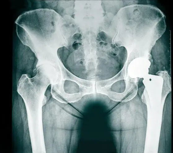
Wear and tear of the hip joint is known as osteoarthritis. This is wear and tear of the joint cartilage, often accompanied by inflammatory processes.
Cause of hip osteoarthritis
The cause of wear and tear can be age-related. However, very often there are also congenital malformations of the hip which, if undetected and untreated, lead to incorrect loading and premature wear. These include, in particular, hip dysplasia (malformation of the acetabulum) or impingement of the hip. Other rheumatologic diseases or circulatory disorders of the femoral head (bone necrosis) can also lead to hip arthrosis.
Typical symptoms of hip osteoarthritis
Indications of advanced osteoarthritis are increasing difficulty getting started and pain during exertion such as long periods of walking. Later, pain may also occur at rest and at night. The pain is typically localized in the groin and/or buttocks as well as at the level of the large tuberosity (outside). It often radiates towards the thigh and knee.
Diagnosis of hip osteoarthritis
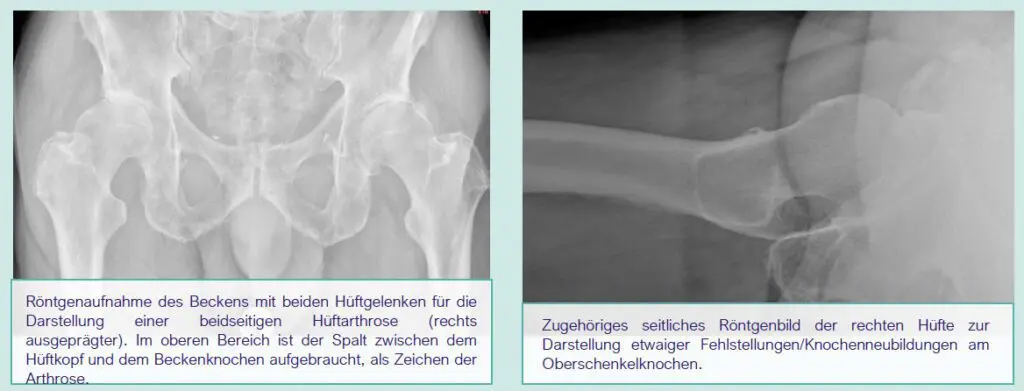
In addition to a medical examination, the most important examination for detecting osteoarthritis is an X-ray of the hip joint.
An overview image of the entire pelvis and a lateral hip image are always taken in order to identify any relevant bone formations, misalignments, etc. that need to be taken into account during treatment. In addition, a reference sphere must be included in the image for surgical planning to ensure correct scaling of the image. If you bring in external X-ray images that do not meet these requirements, we will take new images.
An MRI examination is only useful for certain questions, but is not routinely required. For robot-assisted hip prosthesis surgery, a computer tomography (CT) scan of the pelvis is also performed for individual three-dimensional surgical planning.
